Baker’s Cyst and Its Comprehensive Management

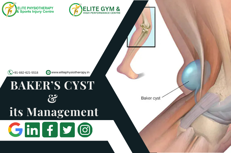
Baker’s cyst, sometimes called a popliteal cyst, is a swelling behind the knee filled with fluid. It happens when synovial fluid builds up in the popliteal bursa, a little sac that lessens friction between the tissues around the knee joint. Although Baker’s cyst is usually not fatal, it can be extremely uncomfortable and restrict physical activity, particularly in athletes and athletic people.

Our specialty at Elite Physiotherapy and Sports Injury Centre is diagnosing and treating Baker’s cysts using advanced modalities and Evidence-based physiotherapy procedures to guarantee the best possible outcome.

Causes of Baker’s Cyst

Usually, an underlying knee condition leads to Baker’s cyst forming. Typical reasons include:

  • Osteoarthritis in the knee
  • Rheumatoid arthritis.
  • Meniscal tears
  • Damage to cartilage
  • Overuse injuries or trauma

For instance, a meniscal tear sustained by an elite athlete who jumps a lot could result in increased production of synovial fluid and the development of a Baker’s cyst.

Mechanism of Injury

When the knee joint becomes inflamed or irritated, too much synovial fluid is produced, which leads to the formation of the cyst. Through a one-way valve, this fluid escapes into the popliteal bursa. The bursa swells and develops into a palpable cyst behind the knee over time.

Signs, Symptoms, and Clinical Features of Baker’s Cyst

The following are Key symptoms of a Baker’s cyst:

  • Tightness and swelling behind the knee
  • Pain that gets worse when you flex your knees or stand for a long time
  • Knee with limited range of motion (ROM)
  • A bulge in the popliteal region that is felt or apparent
  • Discomfort or stiffness during exercising

In extreme situations, the cyst may burst, allowing fluid to seep into the calf and producing symptoms similar to deep vein thrombosis (DVT), including redness, swelling, and warmth.

Diagnostic Methods at Elite Physiotherapy

At Elite Physiotherapy and Sports Injury Centre, we employ a combination of physical and functional assessments to diagnose Baker’s cysts effectively.

Physical Assessment:

  • Check for obvious edema in the popliteal area.
  • palpation to determine the cyst’s presence and size.

Special Physiotherapy Tests for Baker’s Cyst:

  • The Bounce Home Test: Evaluates the knee joint’s integrity and detects any mechanical obstruction or effusion.
  • McMurray’s Test: Assesses meniscal damage that could be a factor in the development of cysts.
  • Apley’s Test: Assists in determining meniscal or ligamentous involvement.
  • Modified Thompson Test: In order to rule out other diseases like DVT, this test looks for fluid leaking into the calf.

Functional Assessment:

  • Examination of Gait patterns.
  • Assessment of joint stability and muscle strength.
  • To identify restrictions, use a range of motion testing.

Physiotherapy Management of Baker’s Cyst at Elite Physiotherapy

We treat Baker’s cysts using an individualized, evidence-based strategy that aims to minimize symptoms, enhance function, and stop recurrence. The cutting-edge treatments we provide are listed below:

1. Electrotherapy Modalities:

  • Shock Wave Therapy: Promotes tissue regeneration, lowers inflammation, and eases popliteal discomfort.
  • The Super Inductive System (SIS): Uses high-frequency electromagnetic stimulation to improve muscle activation and lessen knee stiffness.
  • High-Intensity Class IV Laser Therapy: Promotes the reabsorption of synovial fluid, reduces inflammation, and speeds up tissue recovery.

2. Hydrotherapy:

  • In a regulated setting, water-based workouts improve muscular strength and mobility while lowering joint stress.

3. Cryotherapy:

  • Localized cold therapy reduces pain and swelling, particularly after physical exercise.

4. Manual Therapy:

  • Soft tissue mobilization to ease constriction around the popliteal region.
  • Joint mobilization to increase knee range of motion.

5. Therapeutic Exercises for Baker’s Cyst:

  • Stretching exercises: Stretches for the hamstrings and calves help release tension in the area surrounding the knee joint.
  • Strengthening Exercises: To improve joint stability, concentrate on your quadriceps, hamstrings, and glutes.
  • Proprioception and Balance Training: Enhancing knee control and preventing more injuries are two benefits of proprioception and balance training.

6. Taping Techniques:

  • Kinesiology taping helps to ensure adequate alignment during exercises and lessens the pressure on the knee joint.

7. Progressive Rehabilitation Program:

  • Designed to meet the needs of each participant, this program aims to gradually restore full function and resume daily activities or sports.

Why Choose Elite Physiotherapy for Baker’s Cyst?

Our team at Elite Physiotherapy and Sports Injury Centre is dedicated to providing top-notch care through individualized treatment plans and state-of-the-art technologies. In addition to providing symptom alleviation, our all-encompassing strategy guarantees the long-term avoidance of Baker’s cysts and related knee disorders. With access to cutting-edge techniques including hydrotherapy, shock wave therapy, and super inductive systems, we offer unparalleled proficiency in treating musculoskeletal disorders and sports injuries.

Speak with one of our experts now to start along the path to pain-free mobility and peak performance. Your recovery is our top concern at Elite Physiotherapy!

Patellofemoral Instability: And its Physiotherapy Management

Incorrect alignment or frequent patella (kneecap) dislocation concerning the femoral groove (trochlea) is a complex problem known as patellofemoral instability. If left untreated, this condition may result in chronic joint degeneration, pain, and functional restrictions. Whether they are athletes, active people, or anybody else coping with patellofemoral issues, our goal at Elite Physiotherapy and Sports Injury Centre is to offer the best care possible.

Anatomy of the Patellofemoral Joint

One of the knee’s essential parts is the patellofemoral joint. When walking, jogging, or kneeling, the patella moves smoothly inside the femur’s trochlear groove. Stability is preserved by:

  • Bony structures: The patella’s form and the depth of the trochlear groove.
  • Soft tissue restraints: The patellar tendon, quadriceps muscles, lateral retinaculum, and medial patellofemoral ligament (MPFL) are examples of soft tissue constraints.

When these components become disorganized or imbalanced, instability may result, which might cause the patella to move laterally or dislocate entirely.

Causes of Patellofemoral Instability

Traumatic Events: Patellar dislocation is frequently caused by direct strikes or twisting injuries.

Anatomical Predispositions:

  • trochlear groove that is shallow.
  • High-riding patella, or patella alta.
  • The Q-angle, or the angle formed by the patellar tendon and quadriceps.

Muscle Imbalances:

  • Weakness or delayed vastus medialis obliquus (VMO) activation.
  • tight lateral structures, such as the lateral retinaculum or iliotibial band.

Ligamentous Laxity: People who have conditions like Ehlers-Danlos syndrome may be more prone to instability in the future.

Recurrent Dislocations: The patella is more vulnerable to instability in the future if it has been dislocated.

Symptoms of Patellofemoral Instability

Patients frequently arrive with:

  1. Knee Pain: Knee pain is mostly anterior and is worse when you squat or climb stairs.
  2. Instability:  An unstable knee feels like it’s “giving way.”
  3. Swelling: Particularly following a first displacement.
  4. Reduced Range of Motion: As a result of mechanical impediments or pain.
  5. Apprehension Sign: Fear or discomfort when the patella is moved laterally is an indication of apprehension.

Diagnosis

At Elite Physiotherapy and Sports Injury Centre, we evaluate patellofemoral instability using a variety of cutting-edge diagnostic techniques:

Clinical Assessment:

  • Palpation to find effusion or discomfort.
  • Special tests such as the J-sign observation, apprehension test, and patellar glide test.

Apart from our assessment, patellar instability can be diagnosed also by:

Imaging:

  • X-rays: To evaluate trochlear morphology and bone alignment.
  • MRI: For assessing soft tissues, such as cartilage degeneration and MPFL integrity.
  • CT scans: To evaluate rotational alignment and measure patellar tilt.

Treatment Options

Non-Surgical Management

The first line of treatment is frequently non-surgical, particularly for people who have had only one dislocation or who have modest instability.

Physiotherapy:

The mainstay of treatment for patellofemoral instability is physiotherapy, which aims to enhance patellar tracking, stabilize the joint, and allow for a full return to function. We use a thorough and sophisticated approach at Elite Physiotherapy & Sports Injury Centre, combining manual therapy, therapeutic exercises, and state-of-the-art modalities.

Key Components of Physiotherapy Management
1. Management of Pain and Inflammation
  • Cryotherapy: Using ice packs to minimize swelling and lessen pain.
  • High-Intensity Laser Therapy (HILT): By promoting cellular regeneration, high-intensity laser therapy (HILT) speeds up tissue healing, lowers inflammation, and offers profound pain relief.
  • Shock Wave Therapy: Shock wave therapy is useful for improving the repair of the soft tissues surrounding the patellofemoral joint and reducing chronic pain.
2. Soft Tissue and Joint Mobilization

Manual Therapy:

  • Soft tissue mobilization: For tight lateral structures like the lateral retinaculum and iliotibial band, manual therapy involves mobilizing soft tissues.
  • patellar mobilizations: To enhance trochlear groove alignment and gliding.

Cupping therapy: improves circulation and eases knee-related muscle strain.

3. Neuromuscular Activation and Muscle Strengthening
  • To improve patellar tracking, the quadriceps should be strengthened, especially the vastus medialis obliquus (VMO).
  • To improve total knee stability, progressive resistance training focuses on the calf, gluteal, and hamstring muscles.
  • deep neuromuscular activation, which enhances muscle recruitment and decreases spasticity, through the use of advanced equipment like the Super Inductive System (SIS).
4. Stretching and Flexibility Training
  • To ease tightness and encourage balanced patellar movement, perform stretches for the lateral structures, quadriceps, hip flexors, and iliotibial band.
  • methods for increasing flexibility, such as Proprioceptive Neuromuscular Facilitation (PNF) stretching.
5. Proprioception and Balance Training
  • To improve neuromuscular coordination and joint proprioception, use stability balls, wobble boards, and balancing pads.
  • Functional exercises and sport-specific drills to enhance dynamic stability during intricate motions.
6. Advanced Physiotherapy Modalities
  • Capacitive-Resistive Energy Transfer (C.R.E.T.) therapy: A non-invasive method that enhances tissue circulation and oxygenation, decreases inflammation, and speeds up recovery.
  • Shock Wave Therapy: Encourages soft tissue healing, increases blood flow, and dissolves calcifications.
  • The Super Inductive System (SIS): Uses high-frequency electromagnetic stimulation to help with pain alleviation and muscular strengthening.
7. Functional Rehabilitation
  • Including lunges, step-ups, and squats while paying close attention to proper patellar monitoring.
  • A gradual return to dynamic exercises, such as jumping and sideways motions, to maintain control and alignment when doing sports-specific duties.
8. Patellar Stabilization Techniques
  • Taping: To offer support and adjust patellar alignment, use McConnell or kinesiology taping techniques.
  • Bracing: Personalized patellar braces to reduce excessive movement and boost self-assurance while engaging in exercises.
9. Patient Education
  • Education on good posture, movement patterns, and biomechanics to reduce excessive knee strain.
  • Recurrence prevention guidelines for safe sports and activity participation.

Surgical Management

Recurrent instability, severe anatomical anomalies, or the failure of conservative therapy may all require surgery. Typical procedures consist of:

  • MPFL Reconstruction: Rebuilding the ligament to stop the patella from moving laterally is known as MPFL reconstruction.
  • Trochleoplasty: When a patient’s trochlea is flat, the trochlear groove is deepened.
  • Tibial Tubercle Transfer: Patellar tendon realignment for proper tracking.
  • Lateral Release: Reducing patellar tilt by cutting tight lateral structures.

When surgery is necessary, Elite Physiotherapy and Sports Injury Centre will work post-operative management to provide a smooth transition from surgery to recovery.

At Elite Physiotherapy and Sports Injury Centre, we incorporate these therapies into a comprehensive treatment program that is tailored to each patient’s specific requirements. Our blend of state-of-the-art modalities and evidence-based practices guarantees the best possible recovery and long-term avoidance of patellofemoral instability.