Introduction
- It can be concerning if neck pain extends to the fingers, arm, or shoulder. Fortunately, with the correct care, many people make a full recovery. One of the best non-surgical methods for easing pain, regaining strength, and facilitating a safe return to everyday activities is cervical radiculopathy physiotherapy. Our team at Elite Physiotherapy & Sports Injury Centre offers patients in Delhi NCR evidence-based rehabilitation programs. Whether you’re looking for physiotherapy, physiotherapy Saket, physiotherapy Delhi, premium physiotherapy, or physiotherapy near me, our individualized rehabilitation programs are designed to treat the underlying cause rather than just cover up symptoms.
- Physiotherapy, in contrast to short-term pain management techniques, aims to improve spinal mobility, lessen nerve irritation, restore muscular strength, correct posture, and stop further occurrences. Early intervention frequently results in quicker healing.
What is Cervical Radiculopathy?
- One or more nerves that exit the cervical spine can become compressed, irritated, or inflamed in cervical radiculopathy. The shoulders, arms, hands, and fingers get these nerves from the neck. Compression disrupts normal nerve function, which can result in pain, tingling, numbness, or weakness in the muscles.
- The ailment is frequently referred to as a “pinched nerve in the neck.” Although this explanation is simple to comprehend, the real issue typically involves alterations surrounding the spinal nerve root. These alterations might happen immediately following trauma or prolonged stress, or they can evolve gradually with aging.
- Certain parts of the upper limb receive feeling from each cervical nerve root. Additionally, each neuron regulates certain muscles. As a result, symptoms differ according on the level of the damaged nerve.
- Excellent results are frequently obtained with cautious treatment. Surgery is only used in some situations where there are severe neurological deficits, compression of the spinal cord, or symptoms that do not go away after extensive rehabilitation.
Understanding the Cervical Spine
- Seven vertebrae, numbered C1 through C7, make up the cervical spine. The head is supported by these vertebrae, which also provide an amazing range of mobility. An intervertebral disc, which serves as a shock absorber, is located between the majority of vertebrae.
- Important structures include:
- Cervical vertebrae
- Intervertebral discs
- Facet joints
- Ligaments
- Muscles
- Neural foramina are tiny holes through which the spinal nerves emerge. Nerve compression may happen when these holes shrink or when surrounding tissues swell.
- Appropriate coordination between muscles, joints, ligaments, and nerves is necessary for healthy movement. Over time, even a small imbalance might put more strain on cervical tissues.
How Cervical Radiculopathy Develops
- When there is less room for a nerve root, the ailment typically manifests. The nerve is under more pressure and normal nerve conduction is disrupted when there is less room.
- Compression may result from:
- Bulging intervertebral discs
- Herniated discs
- Bone spurs
- Degenerative arthritis
- Thickened ligaments
- Joint inflammation
- Traumatic injury
- Pain sensitivity is further increased by inflammatory substances secreted near the pinched nerve. As a result, symptoms could last even after the original mechanical compression subsides.
- Patients may progressively experience muscle weakness, diminished coordination, and difficulties in daily activities if they are not well managed.
Causes of Cervical Radiculopathy
- Cervical Disc Herniation
- Cervical Spondylosis
- Foraminal Stenosis
- Degenerative Disc Disease
- Acute Trauma
- Poor Posture
- Repetitive Occupational Stress
- Sports-Related Causes
Signs and Symptoms
- Neck pain that radiates into the shoulder, arm, forearm, or hand
- Sharp, shooting, or electric shock-like pain
- Burning pain along the arm
- Tingling sensation (pins and needles)
- Numbness in specific fingers
- Muscle weakness in the shoulder or arm
- Reduced grip strength
- Difficulty lifting objects
- Pain while turning the head
- Increased pain during coughing or sneezing
- Morning neck stiffness
- Muscle tightness around the neck and shoulder
- Reduced cervical range of motion
- Arm fatigue during prolonged activities
- Difficulty performing overhead work
- Reduced hand coordination in severe cases
When Should You Seek Immediate Medical Attention?
- Sudden loss of arm strength
- Progressive muscle weakness
- Difficulty walking
- Loss of balance
- Problems with bowel or bladder control
- Severe trauma
- Persistent night pain
- Fever with neck pain
- Unexplained weight loss
- Signs of spinal cord compression
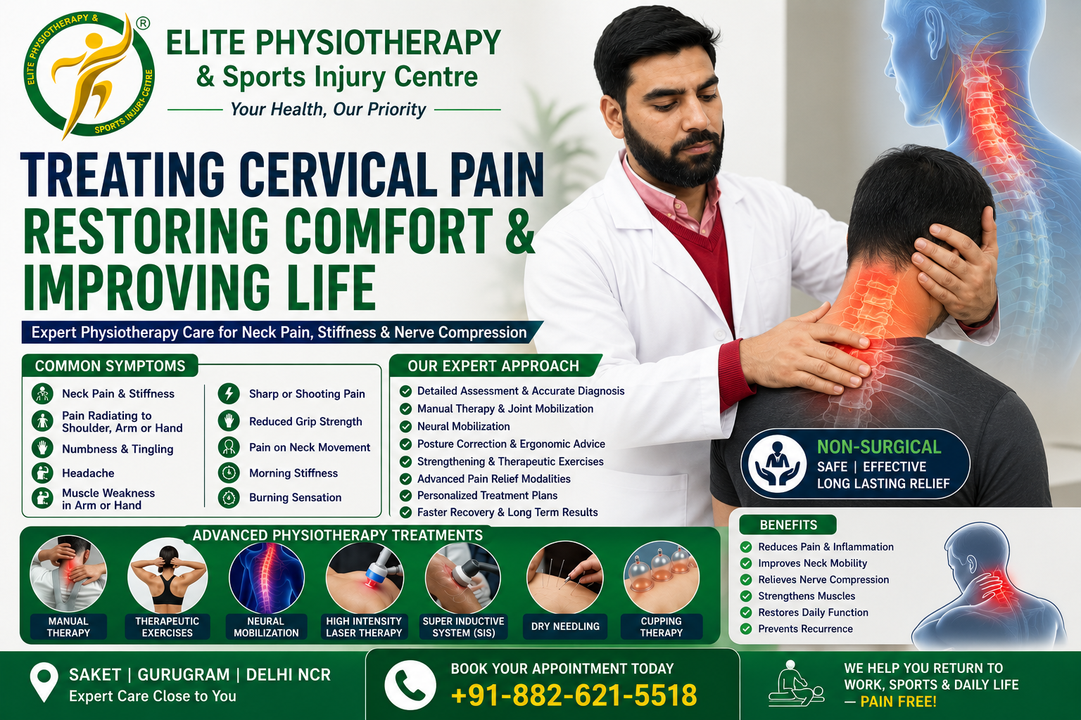
Physiotherapy Management of Cervical Radiculopathy
- Pain Management During the Early Stage
- Avoid prolonged neck flexion
- Avoid repeated overhead activities
- Limit heavy lifting temporarily
- Maintain gentle neck movement
- Avoid complete bed rest
- Change posture regularly
- Use appropriate neck support during prolonged sitting
- Postural Correction
- Patients receive individualized correction strategies for:
- Sitting posture
- Standing posture
- Mobile phone use
- Computer workstations
- Driving posture
- Reading posture
- Sleeping posture
- Manual Therapy
- Cervical joint mobilization
- Thoracic spine mobilization
- Rib mobilization
- Soft tissue mobilization
- Myofascial release
- Muscle energy techniques
- Gentle cervical traction
- Scapular mobilization
- Therapeutic Exercise
- Deep Cervical Flexor Training
- Cervical Range of Motion Exercises
- Scapular Stabilization Exercises
- Shoulder Strengthening
- Upper Limb Strengthening
- Neural Mobilization
- Median nerve glides
- Ulnar nerve glides
- Radial nerve glides
- Cervical nerve mobilization
- Cervical Traction
- Soft Tissue Management
- Ergonomic Training
- Functional Rehabilitation
- Sport-Specific Rehabilitation
- Home Exercise Program
- Activity Modification
- Prevention of Recurrence
Patients are advised to:
- Maintain good posture
- Exercise regularly
- Strengthen neck and shoulder muscles
- Improve thoracic mobility
- Warm up before sports
- Avoid prolonged static positions
- Maintain a healthy body weight
- Use proper lifting techniques
- Schedule regular movement breaks during work
Advanced Physiotherapy Modalities
- High-Intensity Laser Therapy (HILT)
- Super Inductive System (SIS)
- Dry Needling
- Cupping Therapy
- Extracorporeal Shock Wave Therapy (ESWT)
Why Choose Elite Physiotherapy & Sports Injury Centre?
- Our Team offers:
- Evidence-based physiotherapy
- Comprehensive musculoskeletal assessment
- Personalized rehabilitation programs
- Sports physiotherapy expertise
- Advanced manual therapy
- Functional rehabilitation
- Return-to-sport rehabilitation
- Postural correction programs
- Ergonomic assessment
- High-Intensity Laser Therapy
- Super Inductive System
- Dry Needling
- Cupping Therapy
Conclusion
- Cervical radiculopathy can have a major impact on daily activities, sports, and employment. For the majority of patients, however, early diagnosis and evidence-based physical therapy can result in great outcomes.
- Pain is lessened, function is restored, and recurrence is avoided with a thorough rehabilitation program that incorporates patient education, manual therapy, therapeutic exercise, neural mobilization, postural correction, and suitable advanced modalities.
- Our team at Elite Physiotherapy & Sports Injury Centre focuses on determining the underlying cause of your symptoms and developing a personalized treatment plan to enable you to resume your regular activities with confidence.