High-Intensity Class 4 Laser Therapy

At Elite Physiotherapy & Sports Injury Centre, we’re dedicated to offering our patients state-of-the-art care that will accelerate their recovery, improve their performance, and reduce their periods of inactivity. High-intensity Class 4 Laser Therapy (HILT) is one of our most sophisticated and successful techniques. This ground-breaking technology is revolutionizing the treatment of long-term conditions and sports injuries, providing athletes and individuals with unmatched outcomes.

What is High-Intensity Class 4 Laser Therapy?

High-Intensity Class 4 Laser Therapy is a non-invasive procedure that stimulates cellular repair and lessens pain by penetrating deeply into the tissues with concentrated light energy. The Class 4 laser has a higher power output than lower-class lasers, which allows it to more efficiently penetrate deeper tissue layers. This makes it the perfect option for managing chronic pain, sports injuries, and the healing process following surgery.

The Science Behind Class 4 Laser Therapy

Class 4 laser therapy’s therapeutic effects stem from a process known as photobiomodulation. When the laser light interacts with cells, it:

  1. Reduces Inflammation: By lowering the synthesis of pro-inflammatory mediators, the laser light helps to lessen swelling and encourage quicker healing.
  2. Boosts Cellular Metabolism: When cells produce more ATP (adenosine triphosphate), they are better equipped to heal damaged tissues.
  3. Enhances Blood Flow: Better microcirculation expedites the healing process by bringing nutrients and oxygen to the injured area.
  4. Enhances Cellular Metabolism: Increased collagen synthesis is necessary for tissue repair because it fortifies and repairs damaged tissues.

Benefits of High-Intensity Class 4 Laser Therapy at Elite Physiotherapy

  1. Fast Pain Relief: By focusing on pain receptors and lowering inflammation, the treatment offers both short-term and long-term pain relief.
  2. Faster Recovery: When compared to conventional techniques, HILT dramatically shortens the recovery period for sprains, muscle tears, and tendinitis.
  3. Drug-free and non-intrusive: This therapy is perfect for sports and provides a safe substitute for drugs and intrusive procedures.
  4. Customizable Treatments: Every patient can receive the best outcomes possible by modifying the Class 4 laser to meet their specific demands.

Applications in Sports and Rehabilitation

High-Intensity Class 4 Laser Therapy is utilized successfully at Elite Physiotherapy and Sports Injury Centre for:

  • Acute and Chronic Injuries: ranging from sprains of the ligaments to overuse injuries like runner’s knee and tennis elbow.
  • Post-Surgical Recovery: Quickening the healing process following orthopedic procedures like ACL replacement.
  • Muscle and Joint Pain: Managing ailments such as frozen shoulder, bursitis, and osteoarthritis.
  • Performance Optimization: Performance optimization is the process of helping top athletes recover and perform at their best.

Why Choose Elite Physiotherapy and Sports Injury Centre?

Our experience and individualized approach make our center unique. We incorporate High-Intensity Class 4 Laser Therapy into comprehensive treatment plans that are customized to meet the specific needs of each client, making us a center for excellence in sports physiotherapy. Our cutting-edge facilities and highly qualified physiotherapists guarantee that you receive top-notch care in a comforting setting.

A Success Story: Transforming Lives Through Laser Therapy

After only a few sessions of Class 4 Laser Therapy, one of our top athletes who had been recovering from a hamstring strain saw impressive results. The athlete returned to training weeks ahead of schedule after experiencing a significant reduction in pain and a return to mobility. This success story is only one of many that demonstrate how successful this cutting-edge therapy is.

Conclusion

High-Intensity Class 4 Laser Therapy is a revolutionary approach to managing and recovering from sports injuries. We at Elite Physiotherapy and Sports Injury Centre are pleased to provide our customers with this cutting-edge technology so they can live pain-free, perform better, and recover more quickly. Get in touch with us right now to discover the life-changing potential of Class 4 Laser Therapy if you’re recovering from an injury or want to speed up your healing.

Make an Appointment Now!
Learn how High-Intensity Class 4 Laser Therapy can improve your performance and recovery. Get in touch with Elite Physiotherapy and Sports Injury Centre to begin your journey to a more active, healthy lifestyle.

Sinding Larsen Johansson Syndrome

A knee overuse injury known as Sinding Larsen Johansson Syndrome (SLJS) is more common in teenagers who do sports that require repetitive stress, such as running and jumping. We at Elite Physiotherapy and Sports Injury Centre are experts in identifying and managing conditions like SLJS, guaranteeing our patients a successful and individualized recovery process.

Cause and Mechanism of Injury

Repetitive tension on the patellar tendon, which connects to the patella’s lower pole (kneecap), causes SLJS. Particularly during times of rapid growth when bones and tendons are expanding at various rates, this ongoing stress causes inflammation and microtrauma. Young athletes playing high-impact sports like basketball are a classic example, since their frequent jumping puts significant strain on their knee extensor mechanism, leading to SLJS.

Signs and Symptoms

Patients with SLJS may experience:

  • Localized anterior knee pain at the patella’s base that gets worse when you run, jump, or climb stairs.
  • Tenderness and swelling near the patellar tendon insertion site.
  • Restricted knee movement as a result of discomfort, which could cause a noticeable limp.
  • Pain upon palpation of the affected area.

Diagnostic Methods at Elite Physiotherapy and Sports Injury Centre

Our comprehensive assessment includes:

  • Detailed Patient History: Compiling a thorough patient history includes learning about the beginning of symptoms, activity levels, and any recent growth surges.
  • Physical Examination: Examining posture, walking, and knee function using particular tests.
  • Functional Assessment: Determining deficiencies by evaluating the knee’s strength, flexibility, and range of motion.

Special Physiotherapy Tests:

  • Clarke’s Test: In order to identify pain suggestive of patellofemoral dysfunction, Clarke’s Test involves applying pressure to the superior aspect of the patella while the patient contracts the quadriceps.
  • Patellar Compression Test: The patellar compression test is used to detect pain related to problems with the patellar tendon by pressing the patella against the femur.

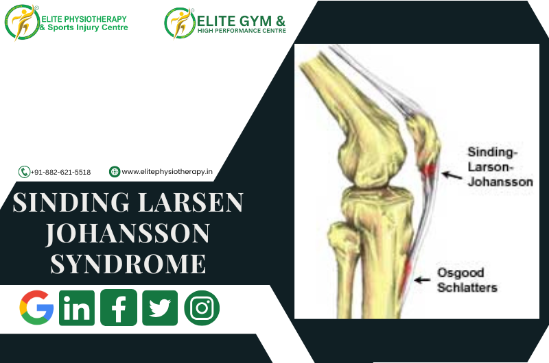
How Osgood-Schlatter disease is different from Sinding-Larsen-Johansson Syndrome

1. Location of the Issue

Osgood-Schlatter Disease (OSD):
Affects the tibial tuberosity, where the patellar tendon inserts on the tibia (shinbone).

Sinding-Larsen-Johansson Syndrome (SLJS):
Involves the inferior pole of the patella, where the patellar tendon attaches to the bottom of the kneecap.

2. Cause and Mechanism

OSD:
Caused by repetitive stress leading to microtrauma at the tibial tuberosity. Excessive pulling by the quadriceps through the patellar tendon results in inflammation and, in some cases, bone fragmentation.

SLJS:
Caused by repetitive traction forces at the inferior pole of the patella, leading to inflammation, microtrauma, or calcification in this area.

3. Clinical Features

OSD Symptoms:

  • Pain and swelling over the tibial tuberosity.
  • Tenderness or prominence at the tibial tuberosity (a bony bump may form).
  • Pain aggravated by running, jumping, or kneeling.

SLJS Symptoms:

  • Pain and tenderness at the inferior pole of the patella.
  • Swelling and discomfort during knee extension activities like squatting or jumping.
  • Pain on palpation directly under the patella.

Physiotherapy Management of Sinding Larsen Johansson Syndrome at Elite Physiotherapy and Sports Injury Centre

Our tailored treatment approach focuses on alleviating pain, reducing inflammation, and restoring function through:

1. Activity Modification: Counseling patients to temporarily stop running and jumping, as these activities aggravate symptoms, in order to promote healing.

2. Cryotherapy: Using ice packs on the afflicted area to reduce pain and swelling.

3. Stretching and Strengthening Exercises:

  • Quadriceps and Hamstring Stretching: Stretching the hamstrings and quadriceps can improve flexibility and lessen patellar tendon strain.
  • Strengthening exercises: To support the knee joint and increase general lower limb strength, concentrate on the quadriceps, hamstrings, and calf muscles.

4. High-End Modalities:

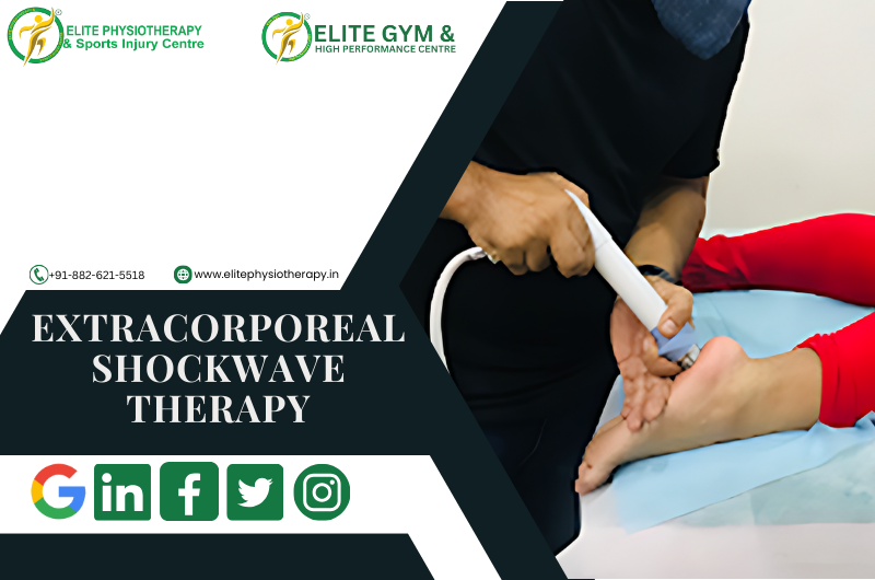
  • Shock Wave Therapy: Using acoustic waves to encourage tissue healing and lessen pain.
  • Capacitive Resistive Energy Transfer (CRET) Therapy: Using electromagnetic radiation to promote tissue regeneration and reduce inflammation is known as capacitive resistive energy transfer, or CRET, therapy.
  • Super Inductive System: The Super Inductive System reduces pain and activates muscles by stimulating neuromuscular regions with high-intensity electromagnetic fields.
  • High-Intensity Class IV Laser Therapy: This method speeds up healing and reduces pain by penetrating deep tissues with laser light.
  • Hydrotherapy: Performing exercises in a warm pool to increase flexibility and strength while lowering joint tension.
  • Cryotherapy: Using cold therapy methods to efficiently treat acute pain and inflammation.

5. Patellar Taping or Bracing: By supporting the patella, patellar taping or bracing lessens the strain on the tendon while performing daily tasks.

6. Patient education: Providing direction on appropriate biomechanics, guidelines for a phased return to sports, and recurrence prevention techniques.

Why to choose Elite Physiotherapy and Sports Injury Centre

Our multidisciplinary team at Elite Physiotherapy and Sports Injury Centre is committed to providing individualized care, utilizing cutting-edge modalities to promote the best possible recovery for those with Sinding-Larsen-Johansson Syndrome. Our dedication to quality guarantees that every patient receives a thorough treatment plan customized to meet their unique requirements, facilitating a prompt and secure return to their preferred activities.

Baker’s Cyst and Its Comprehensive Management

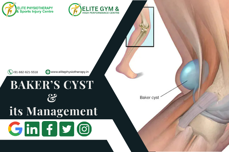
Baker’s cyst, sometimes called a popliteal cyst, is a swelling behind the knee filled with fluid. It happens when synovial fluid builds up in the popliteal bursa, a little sac that lessens friction between the tissues around the knee joint. Although Baker’s cyst is usually not fatal, it can be extremely uncomfortable and restrict physical activity, particularly in athletes and athletic people.

Our specialty at Elite Physiotherapy and Sports Injury Centre is diagnosing and treating Baker’s cysts using advanced modalities and Evidence-based physiotherapy procedures to guarantee the best possible outcome.

Causes of Baker’s Cyst

Usually, an underlying knee condition leads to Baker’s cyst forming. Typical reasons include:

  • Osteoarthritis in the knee
  • Rheumatoid arthritis.
  • Meniscal tears
  • Damage to cartilage
  • Overuse injuries or trauma

For instance, a meniscal tear sustained by an elite athlete who jumps a lot could result in increased production of synovial fluid and the development of a Baker’s cyst.

Mechanism of Injury

When the knee joint becomes inflamed or irritated, too much synovial fluid is produced, which leads to the formation of the cyst. Through a one-way valve, this fluid escapes into the popliteal bursa. The bursa swells and develops into a palpable cyst behind the knee over time.

Signs, Symptoms, and Clinical Features of Baker’s Cyst

The following are Key symptoms of a Baker’s cyst:

  • Tightness and swelling behind the knee
  • Pain that gets worse when you flex your knees or stand for a long time
  • Knee with limited range of motion (ROM)
  • A bulge in the popliteal region that is felt or apparent
  • Discomfort or stiffness during exercising

In extreme situations, the cyst may burst, allowing fluid to seep into the calf and producing symptoms similar to deep vein thrombosis (DVT), including redness, swelling, and warmth.

Diagnostic Methods at Elite Physiotherapy

At Elite Physiotherapy and Sports Injury Centre, we employ a combination of physical and functional assessments to diagnose Baker’s cysts effectively.

Physical Assessment:

  • Check for obvious edema in the popliteal area.
  • palpation to determine the cyst’s presence and size.

Special Physiotherapy Tests for Baker’s Cyst:

  • The Bounce Home Test: Evaluates the knee joint’s integrity and detects any mechanical obstruction or effusion.
  • McMurray’s Test: Assesses meniscal damage that could be a factor in the development of cysts.
  • Apley’s Test: Assists in determining meniscal or ligamentous involvement.
  • Modified Thompson Test: In order to rule out other diseases like DVT, this test looks for fluid leaking into the calf.

Functional Assessment:

  • Examination of Gait patterns.
  • Assessment of joint stability and muscle strength.
  • To identify restrictions, use a range of motion testing.

Physiotherapy Management of Baker’s Cyst at Elite Physiotherapy

We treat Baker’s cysts using an individualized, evidence-based strategy that aims to minimize symptoms, enhance function, and stop recurrence. The cutting-edge treatments we provide are listed below:

1. Electrotherapy Modalities:

  • Shock Wave Therapy: Promotes tissue regeneration, lowers inflammation, and eases popliteal discomfort.
  • The Super Inductive System (SIS): Uses high-frequency electromagnetic stimulation to improve muscle activation and lessen knee stiffness.
  • High-Intensity Class IV Laser Therapy: Promotes the reabsorption of synovial fluid, reduces inflammation, and speeds up tissue recovery.

2. Hydrotherapy:

  • In a regulated setting, water-based workouts improve muscular strength and mobility while lowering joint stress.

3. Cryotherapy:

  • Localized cold therapy reduces pain and swelling, particularly after physical exercise.

4. Manual Therapy:

  • Soft tissue mobilization to ease constriction around the popliteal region.
  • Joint mobilization to increase knee range of motion.

5. Therapeutic Exercises for Baker’s Cyst:

  • Stretching exercises: Stretches for the hamstrings and calves help release tension in the area surrounding the knee joint.
  • Strengthening Exercises: To improve joint stability, concentrate on your quadriceps, hamstrings, and glutes.
  • Proprioception and Balance Training: Enhancing knee control and preventing more injuries are two benefits of proprioception and balance training.

6. Taping Techniques:

  • Kinesiology taping helps to ensure adequate alignment during exercises and lessens the pressure on the knee joint.

7. Progressive Rehabilitation Program:

  • Designed to meet the needs of each participant, this program aims to gradually restore full function and resume daily activities or sports.

Why Choose Elite Physiotherapy for Baker’s Cyst?

Our team at Elite Physiotherapy and Sports Injury Centre is dedicated to providing top-notch care through individualized treatment plans and state-of-the-art technologies. In addition to providing symptom alleviation, our all-encompassing strategy guarantees the long-term avoidance of Baker’s cysts and related knee disorders. With access to cutting-edge techniques including hydrotherapy, shock wave therapy, and super inductive systems, we offer unparalleled proficiency in treating musculoskeletal disorders and sports injuries.

Speak with one of our experts now to start along the path to pain-free mobility and peak performance. Your recovery is our top concern at Elite Physiotherapy!

IT Band Syndrome and Its Management

IT Band Syndrome, also known as Iliotibial Band Syndrome, is a common overuse injury that affects athletes, especially runners, bikers, and those who engage in repetitive lower-limb exercises. We at Elite Physiotherapy and Sports Injury Centre are experts at identifying and successfully treating this illness, enabling athletes to resume their best performance.

Causes of IT Band Syndrome

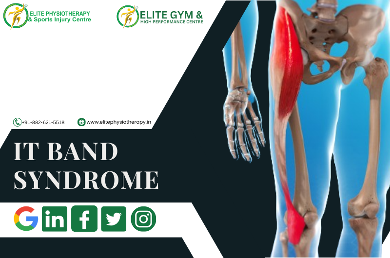
The iliotibial (IT) band is a thick band of connective tissue extending from the hip to the shinbone along the outside of the thigh is called the iliotibial (IT) band. When this band irritates or inflames as a result of excessive contact against the lateral femoral condyle, it can lead to IT Band Syndrome. Typical reasons include:

  • Overuse: Running, cycling, and rowing are examples of repetitive knee flexion and extension exercises.
  • Biomechanical imbalances: Include differences in leg length, weak hip abductors, or poor foot arch mechanics.
  • Inappropriate training methods: Include abrupt increases in mileage, effort, or footwear.
  • Tight IT Band or Surrounding Muscles: The glutes, quadriceps, or hamstrings are less flexible when the IT band or surrounding muscles are tight.

Mechanism of Injury

IT Band Syndrome can result from both on-field and off-field activity. For example, because of insufficient hip stability, a sprinter pushing off quickly during a race may overload their IT band, resulting in microtrauma and ultimately inflammation. Similarly, the IT band may rub too much against the femur if you cycle repeatedly while sitting at an incorrect height.

Signs, Symptoms, and Clinical Features

People with IT Band Syndrome frequently exhibit the following symptoms and indicators:

  • Running or cycling exacerbates lateral knee pain, which is usually acute or burning.
  • Sensitivity along the IT band, especially in the area next to the knee.
  • Localized inflammation or swelling on the knee’s lateral side.
  • Discomfort when sitting for extended periods of time or when going downstairs.
  • When moving, there is an audible snapping or popping sound on the outside of the knee.

Diagnostic Methods at Elite Physiotherapy

To identify the underlying cause of IT Band Syndrome, our diagnostic method combines specific physiotherapy tests with thorough physical and functional evaluations:

1. Physical Assessment:

  • Examination of posture and gait.
  • Palpation along the IT band to detect discomfort.
  • Assessing the hip, knee, and ankle joints’ range of motion and flexibility.

2. Functional Assessment:

  • Evaluation of the mechanics of cycling or running.
  • Hip abductors, glutes, and quadriceps strength tests.

3. Special Physiotherapy Tests:

  • Ober’s Test: Assesses the tightness of the IT band.
  • Noble Compression Test: When knee flexion and extension are performed, the Noble Compression Test detects discomfort or crepitus at the lateral femoral condyle.
  • Modified Thomas Test: The modified Thomas test evaluates hip flexor and IT band flexibility.

Physiotherapy Management at Elite Physiotherapy

We at Elite Physiotherapy and Sports Injury Centre provide a thorough physiotherapy technique that is customized to meet the needs of every patient. In addition to treating underlying biomechanical abnormalities and symptom relief, we also aim to avoid recurrence. This is how we go about it:

1. Pain Management:

  • Cryotherapy: Applying ice to alleviate pain and inflammation is known as cryotherapy.
  • Electrotherapy: Techniques such as TENS or ultrasound that aid in pain management and recovery.

2. Manual Therapy:

  • Soft Tissue Mobilization: Deep tissue massage to relieve tensor fasciae latae (TFL), glutes, and IT band tension.
  • Myofascial Release: Targeting trigger points along the IT band and surrounding muscles is known as myofascial release.
  • Joint Mobilization: Enhancing the ankle, knee, and hip joints’ alignment and range of motion is known as joint mobilization.

3. Stretching Exercises:

  • Stretches that increase the flexibility of the quadriceps, hamstrings, glutes, and IT band can be both dynamic and static.
  • Examples include: The standing hip flexor stretch, pigeon position, and the cross-body IT band stretch.

4. Strengthening Exercises:

Targeted workouts to improve stability and address muscular imbalances:

  • Clamshells: To strengthen the gluteus medius, use clamshells.
  • Lateral Band Walks: Strengthens the hip abductors with lateral band walks.
  • Single-Leg Deadlifts: Improves synergy between the glutes and hamstrings.
  • Step-ups: Increase the general strength and stability of the lower limbs.

5. Biomechanical Correction:

  • Postural corrections and gait retraining are used to maximize movement patterns.
  • Suggestions for appropriate footwear or, if required, orthotics.

6. Return-to-Sport Training:

  • Utilizing sport-specific drills to gradually resume running, cycling, or other sports.
  • Exercises involving plyometry and agility can improve performance while lowering the risk of injury.

Example: IT Band Syndrome in a Cyclist

Think of a professional cyclist who gets lateral knee pain when training hard. We would evaluate their pedaling mechanics and bike fit at Elite Physiotherapy. In addition to physiotherapy treatments, we would address concerns such as incorrect saddle height or cleat position. The cyclist can resume training pain-free and more effectively with the use of focused manual treatment, stretching, and strengthening exercises.

Why Choose Elite Physiotherapy?

At Elite Physiotherapy and Sports Injury Centre, we combine clinical expertise with state-of-the-art facilities to deliver personalized care. Whether you’re a professional athlete or a recreational enthusiast, our evidence-based approach ensures effective recovery and optimal performance. Visit us to experience the best in sports injury management!

PCL Injuries and Advanced Physiotherapy Management

Limiting the tibia’s excessive posterior translation with respect to the femur makes the posterior cruciate ligament (PCL) essential for knee stability. Despite being less frequently injured than the anterior cruciate ligament (ACL), PCL injuries can majorly impact knee function, particularly in sports and physically active people. To ensure the best possible rehabilitation and return to peak performance, we at Elite Physiotherapy & Sports Injury Centre specialize in evidence-based management of PCL injuries.

Causes and Mechanisms of PCL Injury

High-impact trauma or abrupt, powerful movements are common causes of PCL injuries. Typical reasons include:

  • Direct Trauma: A direct impact, such as striking the dashboard in an automobile accident, forces the anterior tibia posteriorly.
  • Sports Injuries: abrupt knee hyperextension or hyperflexion, which is typical in sports like basketball, football, and wrestling.

Example: Sports-Related Mechanism of Injury

A professional football player may land on a flexed knee while simultaneously feeling an opposing force on the shin when diving to rescue a ball. The PCL is overstressed by this hyperflexion and posterior tibial translation, which can result in a partial or total tear.

Signs, Symptoms, and Clinical Features

Individuals with a PCL injury may present with:

  • Mild to moderate stiffness and swelling.
  • Weight-bearing difficulties, particularly when engaging in knee-flexion activities.
  • A sensation of the knee “giving way” or becoming unstable.
  • Knee discomfort in the back, especially when squatting or going downstairs.
  • Loss of full range of motion.

Diagnostic Methods: Physical and Functional Assessment

At Elite Physiotherapy & Sports Injury Centre, we diagnose PCL issues using a thorough and methodical process. Our functional and physical evaluation consists of:

Patient History: A detailed account of the symptoms and mechanism of the injury.
Physical examination: Keeping an eye out for any indications of deformity, edema, or bruises.

Special Physiotherapy Tests:

  • Posterior Drawer Test:  measures the tibia’s posterior translation to evaluate the PCL’s integrity.
  • Sag Sign: When the knee is flexed to 90 degrees, it shows a posterior sagging of the tibia.
  • Quadriceps Active Test: When using the quadriceps, the Quadriceps Active Test measures the posterior tibial displacement.

Our specialists can determine the extent of the injury by combining these tests with functional evaluations.

Physiotherapy Management for PCL Injuries

Our approach to treatment at Elite Physiotherapy & Sports Injury Centre is centered on individualized, non-invasive rehabilitation that is catered to each patient’s need. Our advanced management of physiotherapy consists of:

1. Pain and Swelling Control

  • Compression and the use of ice packs to lessen inflammation.
  • Application of electrical techniques for tissue repair and pain management, such as ultrasonography or interferential treatment (IFT).

2. Joint Mobility and Range of Motion (ROM)

  • Activities with controlled passive and active assistance to regain joint mobility without putting undue strain on the ligament.
  • Utilizing joint mobilizations and other manual therapy procedures to restore mobility.

3. Strengthening and Muscle Activation

  • To improve knee stability and resist posterior tibial translation, strengthen your quadriceps.
  • Exercise your hamstrings carefully to prevent posterior strain on the PCL.
  • To enhance general biomechanics, use progressive resistance training for the hip and core muscles.

4. Neuromuscular Training

  • To regain dynamic stability, perform proprioceptive exercises with balancing boards, BOSU balls, or single-leg stance exercises.
  • Functional training adapted to the patient’s daily routine or sport.

5. Functional and Sport-Specific Rehabilitation

  • For athletes getting ready to resume play, plyometric drills, agility training, and sport-specific activities are recommended.
  • Introduction of pivoting and high-impact activities gradually under the guidance of experts.

5. Advanced Modalities

  • In chronic situations, dry needling or extracorporeal shockwave therapy (ESWT) are used to promote recovery.
  • Blood Flow Restriction (BFR) therapy reduces joint strain while providing effective strengthening.

Why Choose Elite Physiotherapy & Sports Injury Centre?

We take great satisfaction in offering state-of-the-art treatment for PCL injuries at Elite Physiotherapy. Modern technology, manual therapy, and a patient-centered rehabilitation strategy are all incorporated into our approach. Our goals are to improve overall performance and stop future incidents in addition to treating the injury.

By choosing Elite Physiotherapy & Sports Injury Centre, you gain access to:

  • Expert Assessment: Using specific physiotherapy tests, extensive diagnostic capabilities are available.
  • Personalized Care Plans: Interventions specifically designed to meet your rehabilitation objectives and activity level target your specific needs.
  • Advanced Techniques: The most recent evidence-based methods for the best results.

Our dedication to quality guarantees the best possible recuperation for athletes and people of all activity levels. For additional information on managing injuries, check out the blog area of our website or contact one of our professionals for a consultation right now. Allow us to assist you in returning to your passions with greater strength than before!

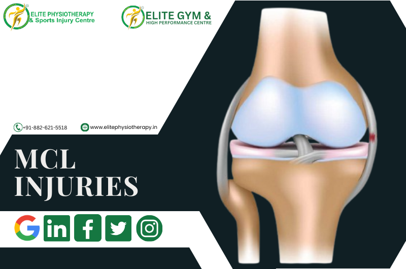
MCL Injuries and its Physiotherapy Management

Our specialty at Elite Physiotherapy and Sports Injury Centre offers evidence-based care to athletes and active people. Among the most frequent knee injuries we treat are medial collateral ligament (MCL) injuries, which frequently arise during sports or other physical activity involving abrupt twisting or impact. This article provides a thorough analysis of MCL injuries, including the causes, diagnostic techniques, and the physiotherapy management procedures we employ to guarantee the best possible outcome.

What is the MCL, and How is it Injured?

On the inside of the knee is a thick, wide ligament called the MCL. It stabilizes the knee against valgus stresses, or inward bending, and joins the femur to the tibia. Usually, MCL injuries are brought on by:

  • Direct Impact: When playing Hockey or football, a strike to the outside of the knee occurs.
  • Abrupt Twisting Motions: Like in skiing or basketball.
  • Overuse: When jogging or cycling, the knee joint experiences repeated stress.

Mechanism of Injury: One common on-field situation that results in MCL tears is when a Football player collides with another player and receives a lateral hit to the knee, causing it to bend inward.

Signs and Symptoms

  • The inside of the knee is tender and painful.
  • Bruising and swelling near the joint.
  • A sensation of looseness or instability in the knee.
  • Inability to fully bend or straighten the leg.
  • Unable to support weight on the injured leg under extreme situations.

Diagnostic Methods of MCL Injuries at Elite Physiotherapy

We use a thorough assessment procedure that consists of:

  • Medical History and Physical Examination: Evaluation of pain, swelling, and valgus instability.
  • Special Physiotherapy Tests: To identify MCL involvement, some tests, such as the Valgus Stress Test, are conducted at 0° and 30° flexion.
  • Functional Mobility Analysis: Assessing knee function during weight-bearing and dynamic motions to spot deficiencies is known as functional mobility analysis.
  • Advanced Imaging (if required): The diagnosis can be verified by working with radiologists to do an MRI or ultrasound.

Comprehensive Physiotherapy Treatment for MCL Injuries at Elite Physiotherapy

Medial Collateral Ligament (MCL) injuries require a systematic, grade-specific treatment approach. Here’s a detailed plan:

1. Phase Acute (First 0–2 Weeks)

Goals:

  • Reduce swelling and pain.
  • Keep the injured ligament safe.

Interventions:

  • Cryotherapy: To reduce inflammation, apply cold packs.
  • Immobilization: To stabilize the joint, use a hinged knee brace.
  • Electrotherapy: Pain is lessened by methods including high-intensity laser therapy and TENS.
  • Gentle ROM Exercises: To preserve knee mobility without putting undue strain on the ligament, engage in passive and active-assisted activities.

2. Subacute Phase (2–6 Weeks)

Goals:

  • Restore muscular activation and range of motion.
  • Start to improve your proprioception and strength.

Interventions:

  • Stretching: Pay attention to your calf, quadriceps, and hamstrings.
  • Isometric Exercises: Work your quadriceps and hamstrings to keep your muscles toned without moving your knees.
  • Proprioception Training: Retrain your balance by using stability aids like wobble boards.
  • Exercises for the Closed Kinetic Chain: Begin with step-ups and mini-squats as tolerated.
  • Manual therapy: Joint alignment is improved by methods such as patellar mobilization.

3. Rehabilitation Phase (6–12 Weeks)

Objectives:

  • Develop dynamic stability and strength.
  • Enhance your functional ability.

Interventions:

  • Incorporate single-leg exercises, resisted lateral walks, and leg presses into your strength training regimen.
  • Dynamic Exercises: Advance to step-downs, lunges, and sideways motions.
  • Neuromuscular Drills: Cutting maneuvers and agility ladders.
  • Plyometrics: To improve power and athletic preparedness, use controlled hops and jumps.
  • Soft Tissue Release: Use myofascial release techniques to relax tense muscles.

4. Advanced Functional Training (12+ Weeks)

Goals:

  • Become prepared for your sport.
  • Prevent re-injury.

Interventions:

  • Speed and Agility Work: Exercises emphasizing rapid direction changes.
  • Sport-Specific Exercises: To regain confidence, mimic game actions.
  • Progressive Load Management: Boost training volume gradually.
  • Functional Testing: To ascertain whether a player is ready to resume play, use strength tests, balance tests, and hop tests.
  • Instruction in Injury Prevention: Provide instruction in appropriate biomechanics, warm-up techniques, and strength-maintenance methods.

Why Choose Elite Physiotherapy for MCL Injuries?

  • Modern Equipment: We include cutting-edge technology in our care, from high-intensity laser therapy to cryotherapy.
  • Holistic Approach: Integrating health and nutrition plans with physical therapy to promote a full recovery.
  • Personalized Care: Every rehabilitation program is customized to the goals, sport, and lifestyle of the individual.

Our goal at Elite Physiotherapy and Sports ailment Centre is to ensure each patient recovers from their injury and regains their strength and self-assurance. To begin your road to recovery if you have an MCL injury, schedule a consultation right now.